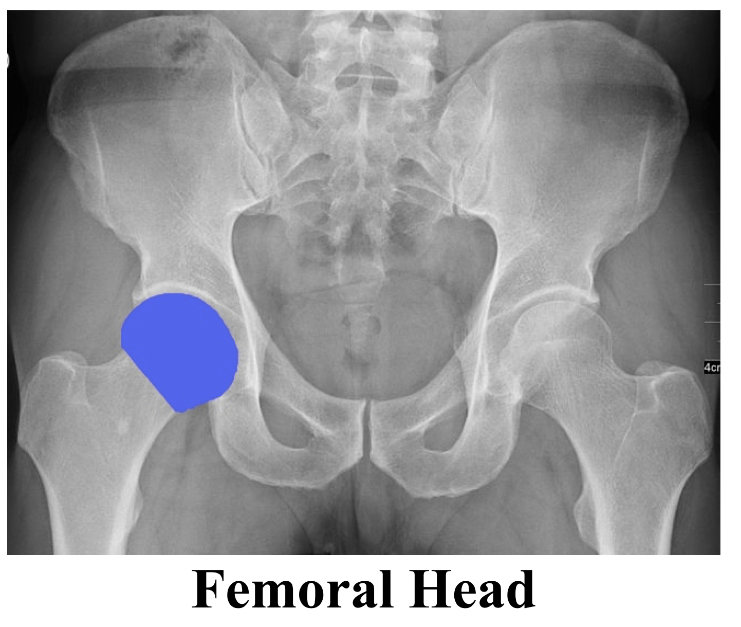
X Ray Pelvis Labelled . 6.1a, b) is a bony ring consisting of paired innominate bones, the sacrum and coccyx. the pelvis series examines the main pelvic ring, obturator foramina, sacroiliac joints, symphysis pubis,. symmetrical greater trochanters + obturator foramen. Too much external rotation of leg. the pelvis radiograph is comprised of the innominate hip bones or os coxae (ischium, pubis and ilium), the sacrum and the proximal femur. No visualization of lesser trochanters. the pelvis (fig.
from savecatchingfire.blogspot.com
the pelvis (fig. 6.1a, b) is a bony ring consisting of paired innominate bones, the sacrum and coccyx. symmetrical greater trochanters + obturator foramen. the pelvis radiograph is comprised of the innominate hip bones or os coxae (ischium, pubis and ilium), the sacrum and the proximal femur. Too much external rotation of leg. the pelvis series examines the main pelvic ring, obturator foramina, sacroiliac joints, symphysis pubis,. No visualization of lesser trochanters.
Pelvic X Ray Anatomy
X Ray Pelvis Labelled 6.1a, b) is a bony ring consisting of paired innominate bones, the sacrum and coccyx. the pelvis radiograph is comprised of the innominate hip bones or os coxae (ischium, pubis and ilium), the sacrum and the proximal femur. the pelvis series examines the main pelvic ring, obturator foramina, sacroiliac joints, symphysis pubis,. Too much external rotation of leg. No visualization of lesser trochanters. symmetrical greater trochanters + obturator foramen. 6.1a, b) is a bony ring consisting of paired innominate bones, the sacrum and coccyx. the pelvis (fig.
From savecatchingfire.blogspot.com
Pelvic X Ray Anatomy X Ray Pelvis Labelled the pelvis (fig. the pelvis series examines the main pelvic ring, obturator foramina, sacroiliac joints, symphysis pubis,. No visualization of lesser trochanters. symmetrical greater trochanters + obturator foramen. the pelvis radiograph is comprised of the innominate hip bones or os coxae (ischium, pubis and ilium), the sacrum and the proximal femur. 6.1a, b) is a bony. X Ray Pelvis Labelled.
From www.pinterest.com.mx
👨🏽💻Want to learn a system for reviewing a pelvic Xray? Read on to X Ray Pelvis Labelled symmetrical greater trochanters + obturator foramen. the pelvis radiograph is comprised of the innominate hip bones or os coxae (ischium, pubis and ilium), the sacrum and the proximal femur. Too much external rotation of leg. the pelvis (fig. the pelvis series examines the main pelvic ring, obturator foramina, sacroiliac joints, symphysis pubis,. 6.1a, b) is a. X Ray Pelvis Labelled.
From quizlet.com
AP Pelvis Radiograph Diagram Diagram Quizlet X Ray Pelvis Labelled 6.1a, b) is a bony ring consisting of paired innominate bones, the sacrum and coccyx. Too much external rotation of leg. symmetrical greater trochanters + obturator foramen. No visualization of lesser trochanters. the pelvis radiograph is comprised of the innominate hip bones or os coxae (ischium, pubis and ilium), the sacrum and the proximal femur. the pelvis. X Ray Pelvis Labelled.
From wikimsk.org
Pelvic Radiograph WikiMSK X Ray Pelvis Labelled No visualization of lesser trochanters. the pelvis radiograph is comprised of the innominate hip bones or os coxae (ischium, pubis and ilium), the sacrum and the proximal femur. 6.1a, b) is a bony ring consisting of paired innominate bones, the sacrum and coccyx. symmetrical greater trochanters + obturator foramen. the pelvis (fig. the pelvis series examines. X Ray Pelvis Labelled.
From www.sciencephoto.com
Female pelvis bones and joints, Xray Stock Image C033/7355 X Ray Pelvis Labelled symmetrical greater trochanters + obturator foramen. the pelvis (fig. the pelvis radiograph is comprised of the innominate hip bones or os coxae (ischium, pubis and ilium), the sacrum and the proximal femur. 6.1a, b) is a bony ring consisting of paired innominate bones, the sacrum and coccyx. No visualization of lesser trochanters. the pelvis series examines. X Ray Pelvis Labelled.
From mavink.com
Pelvic X Ray Labeled X Ray Pelvis Labelled Too much external rotation of leg. 6.1a, b) is a bony ring consisting of paired innominate bones, the sacrum and coccyx. symmetrical greater trochanters + obturator foramen. No visualization of lesser trochanters. the pelvis radiograph is comprised of the innominate hip bones or os coxae (ischium, pubis and ilium), the sacrum and the proximal femur. the pelvis. X Ray Pelvis Labelled.
From mavink.com
Ap Pelvis X Ray Labeled X Ray Pelvis Labelled the pelvis (fig. Too much external rotation of leg. the pelvis series examines the main pelvic ring, obturator foramina, sacroiliac joints, symphysis pubis,. the pelvis radiograph is comprised of the innominate hip bones or os coxae (ischium, pubis and ilium), the sacrum and the proximal femur. symmetrical greater trochanters + obturator foramen. No visualization of lesser. X Ray Pelvis Labelled.
From mavink.com
Pelvic X Ray Labeled X Ray Pelvis Labelled the pelvis radiograph is comprised of the innominate hip bones or os coxae (ischium, pubis and ilium), the sacrum and the proximal femur. the pelvis series examines the main pelvic ring, obturator foramina, sacroiliac joints, symphysis pubis,. No visualization of lesser trochanters. 6.1a, b) is a bony ring consisting of paired innominate bones, the sacrum and coccyx. . X Ray Pelvis Labelled.
From mungfali.com
Pelvis X Ray Labelled X Ray Pelvis Labelled 6.1a, b) is a bony ring consisting of paired innominate bones, the sacrum and coccyx. the pelvis (fig. No visualization of lesser trochanters. symmetrical greater trochanters + obturator foramen. the pelvis radiograph is comprised of the innominate hip bones or os coxae (ischium, pubis and ilium), the sacrum and the proximal femur. the pelvis series examines. X Ray Pelvis Labelled.
From quizlet.com
AP Pelvis XRay Anatomy Diagram Quizlet X Ray Pelvis Labelled Too much external rotation of leg. symmetrical greater trochanters + obturator foramen. the pelvis series examines the main pelvic ring, obturator foramina, sacroiliac joints, symphysis pubis,. 6.1a, b) is a bony ring consisting of paired innominate bones, the sacrum and coccyx. the pelvis (fig. No visualization of lesser trochanters. the pelvis radiograph is comprised of the. X Ray Pelvis Labelled.
From www.pinterest.com.mx
👨🏽💻Want to learn a system for reviewing a pelvic Xray? Read on to X Ray Pelvis Labelled No visualization of lesser trochanters. the pelvis radiograph is comprised of the innominate hip bones or os coxae (ischium, pubis and ilium), the sacrum and the proximal femur. Too much external rotation of leg. 6.1a, b) is a bony ring consisting of paired innominate bones, the sacrum and coccyx. the pelvis (fig. symmetrical greater trochanters + obturator. X Ray Pelvis Labelled.
From savecatchingfire.blogspot.com
Pelvic X Ray Anatomy X Ray Pelvis Labelled 6.1a, b) is a bony ring consisting of paired innominate bones, the sacrum and coccyx. the pelvis (fig. the pelvis radiograph is comprised of the innominate hip bones or os coxae (ischium, pubis and ilium), the sacrum and the proximal femur. No visualization of lesser trochanters. the pelvis series examines the main pelvic ring, obturator foramina, sacroiliac. X Ray Pelvis Labelled.
From www.alamy.com
Pelvis xray front or anterior view. Osteology of the human skeleton X Ray Pelvis Labelled Too much external rotation of leg. the pelvis radiograph is comprised of the innominate hip bones or os coxae (ischium, pubis and ilium), the sacrum and the proximal femur. symmetrical greater trochanters + obturator foramen. the pelvis series examines the main pelvic ring, obturator foramina, sacroiliac joints, symphysis pubis,. the pelvis (fig. 6.1a, b) is a. X Ray Pelvis Labelled.
From mungfali.com
Pelvis X Ray Labelled X Ray Pelvis Labelled the pelvis series examines the main pelvic ring, obturator foramina, sacroiliac joints, symphysis pubis,. the pelvis (fig. symmetrical greater trochanters + obturator foramen. Too much external rotation of leg. the pelvis radiograph is comprised of the innominate hip bones or os coxae (ischium, pubis and ilium), the sacrum and the proximal femur. No visualization of lesser. X Ray Pelvis Labelled.
From gbu-presnenskij.ru
Labeled Hip XRay Anatomy By Naveen Sharma TheRadiologist , 43 OFF X Ray Pelvis Labelled Too much external rotation of leg. 6.1a, b) is a bony ring consisting of paired innominate bones, the sacrum and coccyx. No visualization of lesser trochanters. symmetrical greater trochanters + obturator foramen. the pelvis series examines the main pelvic ring, obturator foramina, sacroiliac joints, symphysis pubis,. the pelvis (fig. the pelvis radiograph is comprised of the. X Ray Pelvis Labelled.
From www.pinterest.com
Pelvis radiograph Medical anatomy, Radiology student, Medical knowledge X Ray Pelvis Labelled the pelvis (fig. symmetrical greater trochanters + obturator foramen. No visualization of lesser trochanters. 6.1a, b) is a bony ring consisting of paired innominate bones, the sacrum and coccyx. the pelvis radiograph is comprised of the innominate hip bones or os coxae (ischium, pubis and ilium), the sacrum and the proximal femur. Too much external rotation of. X Ray Pelvis Labelled.
From www.pinterest.com
Male versus Female Pelvis Labeled Radiographic Anatomy Pelvis X Ray Pelvis Labelled symmetrical greater trochanters + obturator foramen. Too much external rotation of leg. the pelvis series examines the main pelvic ring, obturator foramina, sacroiliac joints, symphysis pubis,. the pelvis (fig. 6.1a, b) is a bony ring consisting of paired innominate bones, the sacrum and coccyx. No visualization of lesser trochanters. the pelvis radiograph is comprised of the. X Ray Pelvis Labelled.
From www.anteriorhipreview.com
normalmalepelvisannotated X Ray Pelvis Labelled No visualization of lesser trochanters. the pelvis series examines the main pelvic ring, obturator foramina, sacroiliac joints, symphysis pubis,. 6.1a, b) is a bony ring consisting of paired innominate bones, the sacrum and coccyx. the pelvis (fig. the pelvis radiograph is comprised of the innominate hip bones or os coxae (ischium, pubis and ilium), the sacrum and. X Ray Pelvis Labelled.